Fundoscopy vs Slit Lamp: The Difference
When comparing fundoscopy vs. slit lamp, the main difference is what they help doctors see: fundoscopy looks deep inside your eye at the retina, while a slit lamp gives a magnified view of your eye’s front parts. A fundus exam uses a special light to check your retina, optic nerve, and blood vessels. A slit lamp uses a bright light and microscope to examine your cornea, iris, lens, and the space between them. Both are vital for eye health checks.
Your eye doctor uses these tools to spot eye diseases early. Fundoscopy is key for conditions like glaucoma, diabetic retinopathy, and macular degeneration. The slit lamp helps diagnose issues such as cataracts, conjunctivitis, and corneal abrasions. Knowing the difference helps you understand your eye exam better and ask your doctor the right questions about your vision health.
- Fundoscopy: Examines the back of your eye (retina, optic nerve).
- Slit Lamp: Examines the front of your eye (cornea, iris, lens).
- Both are non-invasive and quick.
- They help detect a wide range of eye conditions.
- They are standard parts of a comprehensive eye exam.
Let’s dive into what each of these eye examination tools does and why they’re important for keeping your vision clear and healthy.
Looking Inside Your Eye: Fundoscopy vs. Slit Lamp Exam
Your eye doctor uses specialized tools to get a good look at your eyes. Two of the most common are fundoscopy and the slit lamp. While both help your doctor check your vision, they look at **different parts of your eye**. Fundoscopy focuses on the back, and a slit lamp examines the front. Understanding this key difference can help you feel more comfortable and informed during your next eye appointment.
Think of it like this: your eye is a bit like a camera. The slit lamp helps your doctor check the “lens” and the “shutter” (the front parts). Fundoscopy is like looking *through* the lens to see the “film” at the back (your retina and optic nerve). We’ll break down what each exam involves so you know exactly what to expect.
What is a Fundoscopy Exam?
Fundoscopy is a way for your eye doctor to look at the **back of your eye**. This area is also called the fundus. It’s where important structures like your retina, optic nerve, and blood vessels are located. These parts are vital for your vision. Any changes here could signal serious eye problems or even other health issues in your body.
During a fundoscopy, your doctor uses a bright light. This light is often delivered through a tool called an ophthalmoscope. Sometimes, they might use special eye drops to make your pupils larger. This makes it easier to get a clear view of the back of your eye. It’s a quick and painless way to get a lot of information.
Why is Fundoscopy Important?
This exam is essential for detecting and monitoring several eye conditions. For example, it’s a primary tool for spotting diabetic retinopathy. This happens when high blood sugar damages the blood vessels in your retina. Early detection is key to preventing vision loss.
Fundoscopy also helps identify glaucoma. This condition damages the optic nerve, often due to high pressure inside the eye. Damage from glaucoma can be permanent if not treated early. Your doctor can see changes in the optic nerve during a fundoscopy. They can also check for signs of macular degeneration, a leading cause of vision loss in older adults.
We found that changes in the blood vessels at the back of your eye can even indicate problems elsewhere. This includes high blood pressure and diabetes (National Eye Institute). So, a fundoscopy isn’t just about your eyes; it can offer clues about your overall health.
What to Expect During Fundoscopy
Your eye doctor will likely ask you to look in different directions. This helps them get a view of all parts of your retina. They might use a handheld ophthalmoscope, which looks a bit like a small flashlight with lenses. Some doctors may use a larger, more powerful microscope on a stand, called a slit lamp, for this part of the exam. If dilating drops are used, your vision might be blurry for a few hours. It’s a good idea to have someone drive you home if you’re getting these drops.
What is a Slit Lamp Exam?
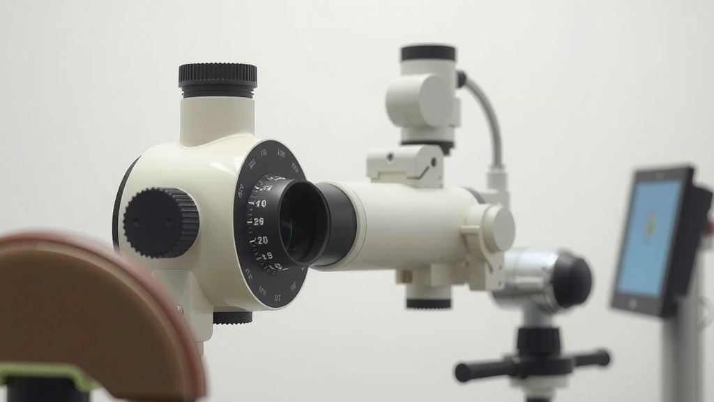
A slit lamp exam gives your doctor a **magnified, detailed view of the front parts of your eye**. This tool combines a powerful light source with a microscope. The light can be adjusted to create a thin beam, like a slit, which illuminates your eye. This allows your doctor to see structures that are too small or clear to see with the naked eye.
The front of your eye includes several important parts. These are the cornea (the clear outer layer), the iris (the colored part), the pupil, the lens (behind the iris), and the space between them. The slit lamp helps your doctor examine each of these very carefully.
Why is a Slit Lamp Exam Important?
This exam is crucial for diagnosing many common eye conditions. For instance, it’s the best way to check for cataracts. A cataract is when the lens of your eye becomes cloudy. The slit lamp allows your doctor to see how cloudy the lens is and how it might be affecting your vision.
If you have red or itchy eyes, a slit lamp can help diagnose conjunctivitis (pink eye). It can also spot minor injuries like corneal abrasions, which are scratches on the surface of your eye. Many doctors also use it to check for signs of dry eye syndrome, where your eyes don’t produce enough tears.
Experts at the Cleveland Clinic highlight that the slit lamp is also used to check the pressure inside your eye for glaucoma and to examine the drainage angle of your eye.
What to Expect During a Slit Lamp Exam
You’ll be asked to place your chin and forehead on rests. This keeps your head still while the doctor looks at your eye. The slit lamp itself won’t touch you. The doctor will move the instrument around your eye. You might see bright lights, but it’s generally not uncomfortable. The entire process is quick, usually taking just a few minutes for each eye.
Comparing Fundoscopy and Slit Lamp Exams
While both exams are vital, they serve distinct purposes. Here’s a quick rundown of their main differences:
| Feature | Fundoscopy | Slit Lamp Exam |
|---|---|---|
| Area Examined | Back of the eye (retina, optic nerve, blood vessels) | Front of the eye (cornea, iris, lens, eyelids) |
| Main Purpose | Detects conditions like diabetic retinopathy, glaucoma, macular degeneration. | Detects conditions like cataracts, conjunctivitis, corneal scratches, dry eye. |
| Tools Used | Ophthalmoscope (handheld or part of slit lamp), dilating drops (sometimes). | Slit Lamp (microscope with adjustable light beam). |
| What You See | A view of your retina’s interior. | A magnified view of your eye’s external and internal front structures. |
Which Exam is More Important?
Neither exam is inherently “more important.” They are both **essential components of a complete eye exam**. Your eye doctor will choose which tools to use based on your specific needs and symptoms. Often, a comprehensive eye exam will include elements of both.
Think of them as different tools in a toolbox. You wouldn’t use a hammer to tighten a screw, right? Similarly, your doctor uses the right tool for the right job. For a full picture of your eye health, both the front and the back need to be checked thoroughly.
Can One Tool Be Used for Both?
Yes, in some cases. Many optometrists and ophthalmologists use the slit lamp to perform fundoscopy. They attach a special lens to the slit lamp that allows them to see the back of your eye with magnification. This is a very common practice and often provides a more detailed view than a handheld ophthalmoscope alone.
However, a dedicated handheld ophthalmoscope is still a portable and effective tool for fundoscopy, especially in settings where a slit lamp might not be available. The key takeaway is the purpose of the examination, not just the specific device.
Checklist: What to Remember About Your Eye Exams
To help you prepare for your next visit, here’s a quick checklist:
- Understand fundoscopy checks the back of your eye.
- Know slit lamp exams look at the front of your eye.
- Both are non-painful and very quick.
- These exams help spot common and serious eye diseases early.
- Ask your doctor if dilating drops are needed for fundoscopy.
- Don’t hesitate to ask your doctor what they are looking for during each exam.
Conclusion
You’ve learned that fundoscopy and slit lamp exams are like two halves of a whole when it comes to checking your eye health. Fundoscopy gives your doctor a look at the vital back of your eye, checking your retina and optic nerve for issues like diabetic retinopathy or glaucoma. The slit lamp provides a magnified view of the front, helping to catch cataracts or corneal scratches. Neither is more important; they work together to give a complete picture. Understanding these differences means you can feel more confident during your next eye appointment. Don’t hesitate to ask your eye doctor about what they’re seeing and why each exam is important for your vision.
Frequently Asked Questions
Do I need both a fundoscopy and a slit lamp exam every time?
Not necessarily. Your eye doctor will decide which exams are best based on your age, overall health, and any symptoms you’re experiencing. Often, a routine eye exam will include aspects of both to ensure all parts of your eye are checked.
Can a slit lamp be used for fundoscopy?
Yes, many eye doctors use the slit lamp with a special lens attachment to perform a fundoscopy. This method can provide a highly magnified and detailed view of the back of your eye, often offering more clarity than a handheld ophthalmoscope alone.
Is a fundoscopy exam painful?
A standard fundoscopy exam is not painful. It involves shining a light into your eye. If dilating eye drops are used to widen your pupils for a better view, you might experience temporary light sensitivity or blurry vision, but this is not painful.
How long does a slit lamp exam take?
A slit lamp exam is very quick. You’ll rest your chin and forehead on the equipment, and the doctor will examine each eye with the magnified light. The entire process usually takes only a few minutes per eye.
What if I wear glasses or contact lenses? Do I need to take them off for these exams?
For a slit lamp exam, you will likely need to remove your glasses but can usually keep your contact lenses in. For fundoscopy, whether you keep your glasses on or off might depend on the specific technique your doctor uses and the clarity of your prescription.


![10 Best Acrylic Nail Lamp [2026]](https://bestdesklamp.com/wp-content/uploads/2026/09/10-best-acrylic-nail-lamp-2026-featured-768x432.jpg)

![10 Best Bedside Lamp With Usb [2026]](https://bestdesklamp.com/wp-content/uploads/2026/08/10-best-bedside-lamp-with-usb-2026-featured-768x432.jpg)